Browse Items (284 total)

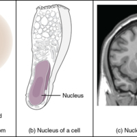
What Is a Nucleus.jpg
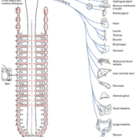
(a) The nucleus of an atom contains its protons and neutrons. (b) The nucleus of a cell is the organelle that contains DNA. (c) A nucleus in the CNS is a localized center of function with the cell bodies of several neurons, shown here circled in red

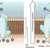
Voltage-Gated Channels.jpg
Voltage-gated channels open when the transmembrane voltage changes around them. Amino acids in the structure of the protein are sensitive to charge and cause the pore to open to the selected ion.

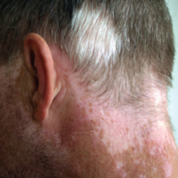
Vitiligo.jpg
Individuals with vitiligo experience depigmentation that results in lighter colored patches of skin. The condition is especially noticeable on darker skin.

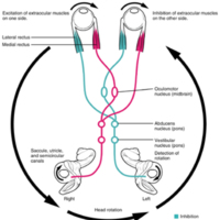
Vestibulo-ocular Reflex.jpg
Connections between the vestibular system and the cranial nerves controlling eye movement keep the eyes centered on a visual stimulus, even though the head is moving. During head movement, the eye muscles move the eyes in the opposite direction as…

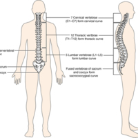
Vertebral Column.jpg
The adult vertebral column consists of 24 vertebrae, plus the sacrum and coccyx. The vertebrae are divided into three regions: cervical C1–C7 vertebrae, thoracic T1–T12 vertebrae, and lumbar L1–L5 vertebrae. The vertebral column is curved, with two…

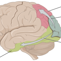
Ventral and Dorsal Visual Streams.jpg
From the primary visual cortex in the occipital lobe, visual processing continues in two streams—one into the temporal lobe and one into the parietal lobe.

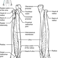
Ulna and Radius.jpg
The ulna is located on the medial side of the forearm, and the radius is on the lateral side. These bones are attached to each other by an interosseous membrane.

UGA Anatomy and Physiology 2 Lab Manual.pdf
The manual contains the following labs:

Blood Composition
Blood Typing
Heart Anatomy
Cardiovascular Physiology
Systemic Blood Vessels
Anatomy of the Respiratory System
Physiology of the Respiratory System

UGA Anatomy and Physiology 1 Lab Manual.pdf
The manual contains the following labs:

Introduction
Tissues
Integument
Introduction to the Skeleton
Axial Skeleton: Skull
Axial Skeleton: Vertebral
Appendicular Skeleton: Introduction and Pectoral Girdle

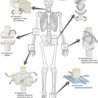
Types of Synovial Joints.jpg
The six types of synovial joints allow the body to move in a variety of ways. (a) Pivot joints allow for rotation around an axis, such as between the first and second cervical vertebrae, which allows for side-to-side rotation of the head. (b) The…

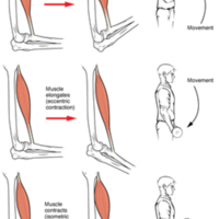
Types of Muscle Contractions.jpg
During isotonic contractions, muscle length changes to move a load. During isometric contractions, muscle length does not change because the load exceeds the tension the muscle can generate.

Types of Fractures.jpg
Compare healthy bone with different types of fractures: (a) closed fracture, (b) open fracture, (c) transverse fracture, (d) spiral fracture, (e) comminuted fracture, (f) impacted fracture, (g) greenstick fracture, and (h) oblique fracture.

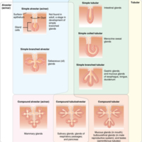
Types of Exocrine Glands.jpg
Exocrine glands are classified by their structure.

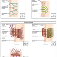
Types of Cell Junctions.jpg
The three basic types of cell-to-cell junctions are tight junctions, gap junctions, and anchoring junctions.

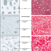
Types of Cartilage.jpg
Cartilage is a connective tissue consisting of collagenous fibers embedded in a firm matrix of chondroitin sulfates. (a) Hyaline cartilage provides support with some flexibility. The example is from dog tissue. (b) Fibrocartilage provides some…

Transmission of Sound Waves to Cochlea.jpg
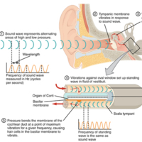
A sound wave causes the tympanic membrane to vibrate. This vibration is amplified as it moves across the malleus, incus, and stapes. The amplified vibration is picked up by the oval window causing pressure waves in the fluid of the scala vestibuli…

Translation from RNA to Protein.jpg
During translation, the mRNA transcript is “read” by a functional complex consisting of the ribosome and tRNA molecules. tRNAs bring the appropriate amino acids in sequence to the growing polypeptide chain by matching their anti-codons with codons on…

Transcription from DNA to mRNA.jpg
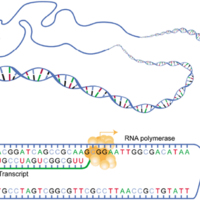
In the first of the two stages of making protein from DNA, a gene on the DNA molecule is transcribed into a complementary mRNA molecule.

Topographic Mapping of the Retina onto the Visual Cortex.jpg
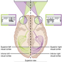
The visual field projects onto the retina through the lenses and falls on the retinae as an inverted, reversed image. The topography of this image is maintained as the visual information travels through the visual pathway to the cortex.

Tissue Membranes.jpg
The two broad categories of tissue membranes in the body are (1) connective tissue membranes, which include synovial membranes, and (2) epithelial membranes, which include mucous membranes, serous membranes, and the cutaneous membrane, in other…

Tibia and Fibula.jpg
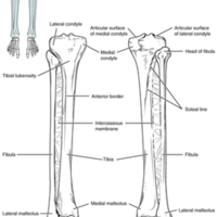
The tibia is the larger, weight-bearing bone located on the medial side of the leg. The fibula is the slender bone of the lateral side of the leg and does not bear weight.

Three Forms of Endocytosis.jpg
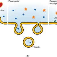
Endocytosis is a form of active transport in which a cell envelopes extracellular materials using its cell membrane. (a) In phagocytosis, which is relatively nonselective, the cell takes in a large particle. (b) In pinocytosis, the cell takes in…

Thoracic Vertebrae.jpg
A typical thoracic vertebra is distinguished by the spinous process, which is long and projects downward to overlap the next inferior vertebra. It also has articulation sites (facets) on the vertebral body and a transverse process for rib attachment.

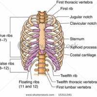
Thoracic Cage.jpg
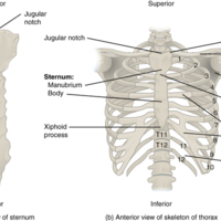
The thoracic cage is formed by the (a) sternum and (b) 12 pairs of ribs with their costal cartilages. The ribs are anchored posteriorly to the 12 thoracic vertebrae. The sternum consists of the manubrium, body, and xiphoid process. The ribs are…

Thermoregulation.jpg
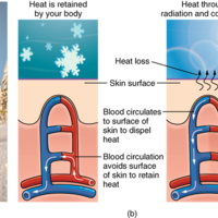
During strenuous physical activities, such as skiing (a) or running (c), the dermal blood vessels dilate and sweat secretion increases (b). These mechanisms prevent the body from overheating. In contrast, the dermal blood vessels constrict to…

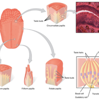
The Tongue.jpg
The tongue is covered with small bumps, called papillae, which contain taste buds that are sensitive to chemicals in ingested food or drink. Different types of papillae are found in different regions of the tongue. The taste buds contain specialized…

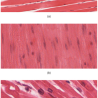
The Three Types of Muscle Tissue.jpg
The body contains three types of muscle tissue: (a) skeletal muscle, (b) smooth muscle, and (c) cardiac muscle.

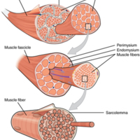
The Three Connective Tissue Layers.jpg
Bundles of muscle fibers, called fascicles, are covered by the perimysium. Muscle fibers are covered by the endomysium.

The Three Components of the Cytoskeleton.jpg
The cytoskeleton consists of (a) microtubules, (b) microfilaments, and (c) intermediate filaments. The cytoskeleton plays an important role in maintaining cell shape and structure, promoting cellular movement, and aiding cell division.

The T-tubule.jpg
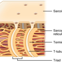
Narrow T-tubules permit the conduction of electrical impulses. The SR functions to regulate intracellular levels of calcium. Two terminal cisternae (where enlarged SR connects to the T-tubule) and one T-tubule comprise a triad—a “threesome” of…

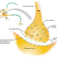
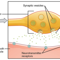
The Synapse.jpg
The synapse is a connection between a neuron and its target cell (which is not necessarily a neuron). The presynaptic element is the synaptic end bulb of the axon where Ca2+ enters the bulb to cause vesicle fusion and neurotransmitter release. The…

The Sliding Filament Model of Muscle Contraction.jpg
When a sarcomere contracts, the Z lines move closer together, and the I band becomes smaller. The A band stays the same width. At full contraction, the thin and thick filaments overlap completely.

The Sensory Input.jpg
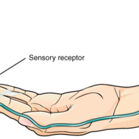
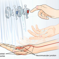
Receptors in the skin sense the temperature of the water.

The Sensory Homunculus.jpg
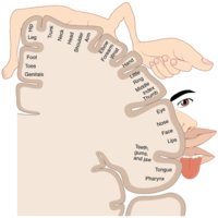
A cartoon representation of the sensory homunculus arranged adjacent to the cortical region in which the processing takes place.

The Sarcomere.jpg
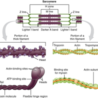
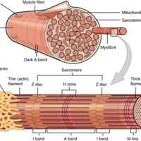
The sarcomere, the region from one Z-line to the next Z-line, is the functional unit of a skeletal muscle fiber.

The Q-Angle.jpg
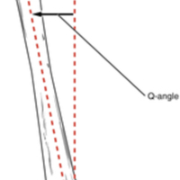
The Q-angle is a measure of the amount of lateral deviation of the femur from the vertical line of the tibia. Adult females have a larger Q-angle due to their wider pelvis than adult males.

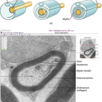
The Process of Myelination.jpg
Myelinating glia wrap several layers of cell membrane around the cell membrane of an axon segment. A single Schwann cell insulates a segment of a peripheral nerve, whereas in the CNS, an oligodendrocyte may provide insulation for a few separate axon…

The Olfactory System.jpg
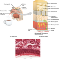
(a) The olfactory system begins in the peripheral structures of the nasal cavity. (b) The olfactory receptor neurons are within the olfactory epithelium. (c) Axons of the olfactory receptor neurons project through the cribriform plate of the ethmoid…

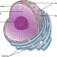
The Nucleus.jpg
The nucleus is the control center of the cell. The nucleus of living cells contains the genetic material that determines the entire structure and function of that cell.

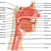
The Nose and its Adjacent Structures.jpg
Several bones that help form the walls of the nasal cavity have air-containing spaces called the paranasal sinuses, which serve to warm and humidify incoming air. Sinuses are lined with a mucosa. Each paranasal sinus is named for its associated bone:…

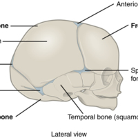
The Newborn Skull.jpg
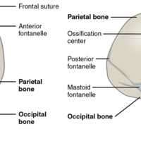
The fontanelles of a newborn’s skull are broad areas of fibrous connective tissue that form fibrous joints between the bones of the skull.

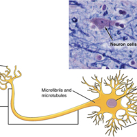
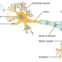
The Neuron.jpg
The cell body of a neuron, also called the soma, contains the nucleus and mitochondria. The dendrites transfer the nerve impulse to the soma. The axon carries the action potential away to another excitable cell.

The Motor Response.jpg
On the basis of the sensory input and the integration in the CNS, a motor response is formulated and executed.

Human Anatomy.jpg
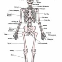
The human skeleton provides shape and form to the human body
Our vital organs in our body are protected by our skeleton. More specifically our brain which is protected by what is called the skull and our heart and lungs are protected by our rib…

The Hip Bone.jpg
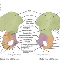
The adult hip bone consists of three regions. The ilium forms the large, fan-shaped superior portion, the ischium forms the posteroinferior portion, and the pubis forms the anteromedial portion.

The Genetic Code.jpg
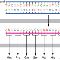
DNA holds all of the genetic information necessary to build a cell’s proteins. The nucleotide sequence of a gene is ultimately translated into an amino acid sequence of the gene’s corresponding protein.

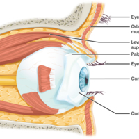
The Eye in the Orbit.jpg
The eye is located within the orbit and surrounded by soft tissues that protect and support its function. The orbit is surrounded by cranial bones of the skull.

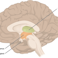
The Diencephalon.jpg
The diencephalon is composed primarily of the thalamus and hypothalamus, which together define the walls of the third ventricle. The thalami are two elongated, ovoid structures on either side of the midline that make contact in the middle. The…

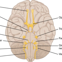
The Cranial Nerves.jpg
The anatomical arrangement of the roots of the cranial nerves observed from an inferior view of the brain.

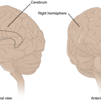
The Cerebrum.jpg
The cerebrum is a large component of the CNS in humans, and the most obvious aspect of it is the folded surface called the cerebral cortex.

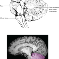
The Cerebellum.jpg
The cerebellum is situated on the posterior surface of the brain stem. Descending input from the cerebellum enters through the large white matter structure of the pons. Ascending input from the periphery and spinal cord enters through the fibers of…

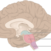
The Brain Stem.jpg
The brain stem comprises three regions: the midbrain, the pons, and the medulla.

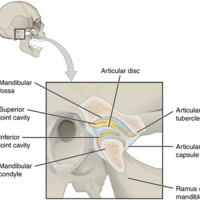
Temporomandibular Joint.jpg
The temporomandibular joint is the articulation between the temporal bone of the skull and the condyle of the mandible, with an articular disc located between these bones. During depression of the mandible (opening of the mouth), the mandibular…

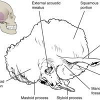
Temporal Bone.jpg
A lateral view of the isolated temporal bone shows the squamous, mastoid, and zygomatic portions of the temporal bone.

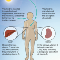
Synthesis of Vitamin D.jpg
Sunlight is one source of vitamin D.

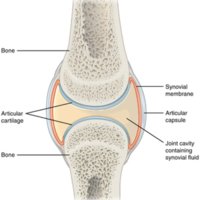
Synovial Joints.jpg
Synovial joints allow for smooth movements between the adjacent bones. The joint is surrounded by an articular capsule that defines a joint cavity filled with synovial fluid. The articulating surfaces of the bones are covered by a thin layer of…

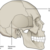
Suture Joints of Skull.jpg
The suture joints of the skull are an example of a synarthrosis, an immobile or essentially immobile joint.

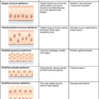
Summary of Epithelial Tissue Cells.jpg
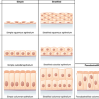
A stratified epithelium consists of several stacked layers of cells. This epithelium protects against physical and chemical wear and tear. The stratified epithelium is named by the shape of the most apical layer of cells, closest to the free space.…

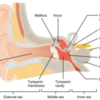
Structures of the Ear.jpg
The external ear contains the auricle, ear canal, and tympanic membrane. The middle ear contains the ossicles and is connected to the pharynx by the Eustachian tube. The inner ear contains the cochlea and vestibule, which are responsible for audition…

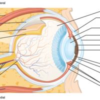
Structure of the Eye.jpg
The sphere of the eye can be divided into anterior and posterior chambers. The wall of the eye is composed of three layers: the fibrous tunic, vascular tunic, and neural tunic. Within the neural tunic is the retina, with three layers of cells and two…

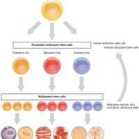
Stem Cells.png
The capacity of stem cells to differentiate into specialized cells make them potentially valuable in therapeutic applications designed to replace damaged cells of different body tissues.

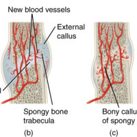
Stages in Fracture Repair.jpg
The healing of a bone fracture follows a series of progressive steps: (a) A fracture hematoma forms. (b) Internal and external calli form. (c) Cartilage of the calli is replaced by trabecular bone. (d) Remodeling occurs.

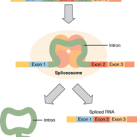
Splicing DNA.jpg
In the nucleus, a structure called a spliceosome cuts out introns (noncoding regions) within a pre-mRNA transcript and reconnects the exons.

Spinal Cord and Root Ganglion.jpg
The slide includes both a cross-section of the lumbar spinal cord and a section of the dorsal root ganglion (see also [link]) (tissue source: canine).

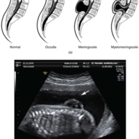
Spinal Bifida.jpg
(a) Spina bifida is a birth defect of the spinal cord caused when the neural tube does not completely close, but the rest of development continues. The result is the emergence of meninges and neural tissue through the vertebral column. (b) Fetal…

Sphenoid Bone.jpg
Shown in isolation in (a) superior and (b) posterior views, the sphenoid bone is a single midline bone that forms the anterior walls and floor of the middle cranial fossa. It has a pair of lesser wings and a pair of greater wings. The sella turcica…

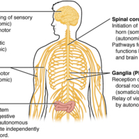
Somatic, Autonomic, and Enteric Structures of the Nervous System.jpg
Somatic structures include the spinal nerves, both motor and sensory fibers, as well as the sensory ganglia (posterior root ganglia and cranial nerve ganglia). Autonomic structures are found in the nerves also, but include the sympathetic and…

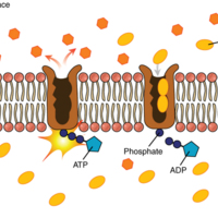
Sodium-Potassium Pump.jpg
The sodium-potassium pump is found in many cell (plasma) membranes. Powered by ATP, the pump moves sodium and potassium ions in opposite directions, each against its concentration gradient. In a single cycle of the pump, three sodium ions are…

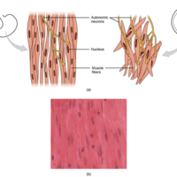
Smooth Muscle Tissue.jpg
Smooth muscle tissue is found around organs in the digestive, respiratory, reproductive tracts and the iris of the eye.

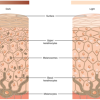
Skin Pigmentation.jpg
The relative coloration of the skin depends of the amount of melanin produced by melanocytes in the stratum basale and taken up by keratinocytes.

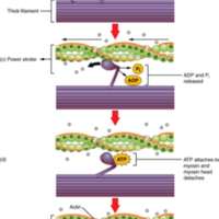
Skeletal Muscle Contraction.jpg
(a) The active site on actin is exposed as calcium binds to troponin. (b) The myosin head is attracted to actin, and myosin binds actin at its actin-binding site, forming the cross-bridge. (c) During the power stroke, the phosphate generated in the…

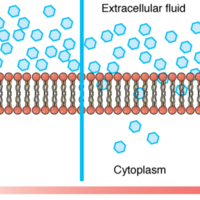
Simple Diffusion across the Cell (Plasma) Membrane.jpg
The structure of the lipid bilayer allows small, uncharged substances such as oxygen and carbon dioxide, and hydrophobic molecules such as lipids, to pass through the cell membrane, down their concentration gradient, by simple diffusion.

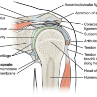
914_Shoulder_Joint.jpg
The shoulder joint is called the glenohumeral joint. This is a ball-and-socket joint formed by the articulation between the head of the humerus and the glenoid cavity of the scapula ([link]). This joint has the largest range of motion of any joint in…

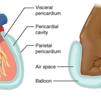
112_Serous_Membrane_new.jpg
A serous membrane (also referred to a serosa) is one of the thin membranes that cover the walls and organs in the thoracic and abdominopelvic cavities. The parietal layers of the membranes line the walls of the body cavity (pariet- refers to a cavity…

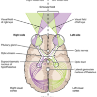
Segregation of Visual Field Information at the Optic Chiasm.jpg
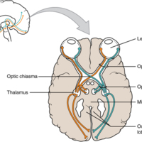
Contralateral visual field information from the lateral retina projects to the ipsilateral brain, whereas ipsilateral visual field information has to decussate at the optic chiasm to reach the opposite side of the brain.

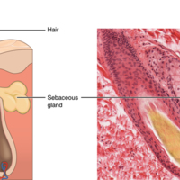
Sebaceous Glands.jpg
These glands secrete oils that lubricate and protect the skin. They are holocrine glands and they are destroyed after releasing their contents. New glandular cells form to replace the cells that are lost

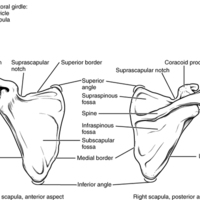
Scapula.jpg
The isolated scapula is shown here from its anterior (deep) side and its posterior (superficial) side.

Sagittal Section of Skull.jpg
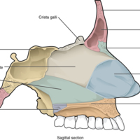
This midline view of the sagittally sectioned skull shows the nasal septum.

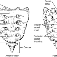
Sacrum and Coccyx.jpg
The sacrum is formed from the fusion of five sacral vertebrae, whose lines of fusion are indicated by the transverse ridges. The fused spinous processes form the median sacral crest, while the lateral sacral crest arises from the fused transverse…

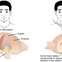
Rotational Coding by Semicircular Canals.jpg
Rotational movement of the head is encoded by the hair cells in the base of the semicircular canals. As one of the canals moves in an arc with the head, the internal fluid moves in the opposite direction, causing the cupula and stereocilia to bend.…

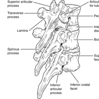
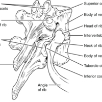
Rib Articulation in Thoracic Vertebrae.jpg
Thoracic vertebrae have superior and inferior articular facets on the vertebral body for articulation with the head of a rib, and a transverse process facet for articulation with the rib tubercle.

Retinal Isomers.jpg
The retinal molecule has two isomers, (a) one before a photon interacts with it and (b) one that is altered through photoisomerization.

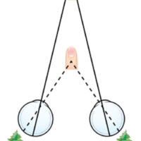
Retinal Disparity.jpg
Because of the interocular distance, which results in objects of different distances falling on different spots of the two retinae, the brain can extract depth perception from the two-dimensional information of the visual field.

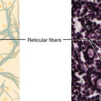
Reticular Tissue.jpg
This is a loose connective tissue made up of a network of reticular fibers that provides a supportive framework for soft organs.

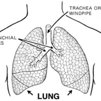
lung_PD_PearsonScottForesman.png
The job of the respiratory system is to take oxygen from the air we breathe and get it to different parts of the body. Our bodies and the cells in them need oxygen (written with the chemical symbol O2) to live. Our cells give off carbon dioxide…

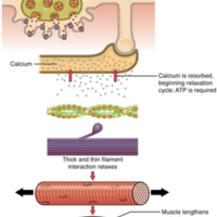
Relaxation of a Muscle Fiber.jpg
Ca++ ions are pumped back into the SR, which causes the tropomyosin to reshield the binding sites on the actin strands. A muscle may also stop contracting when it runs out of ATP and becomes fatigued.

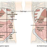
111_Abdominal_Quadrant_Regions.jpg
The more detailed regional approach subdivides the cavity with one horizontal line immediately inferior to the ribs and one immediately superior to the pelvis, and two vertical lines drawn as if dropped from the midpoint of each clavicle…

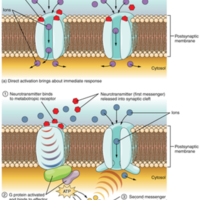
Receptor Types.jpg
(a) An ionotropic receptor is a channel that opens when the neurotransmitter binds to it. (b) A metabotropic receptor is a complex that causes metabolic changes in the cell when the neurotransmitter binds to it (1). After binding, the G protein…

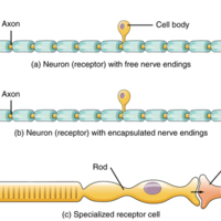
Receptor Classification by Cell Type.jpg
Receptor cell types can be classified on the basis of their structure. Sensory neurons can have either (a) free nerve endings or (b) encapsulated endings. Photoreceptors in the eyes, such as rod cells, are examples of (c) specialized receptor cells.…

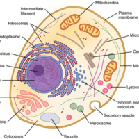
Prototypical Human Cell.jpg
While this image is not indicative of any one particular human cell, it is a prototypical example of a cell containing the primary organelles and internal structures.

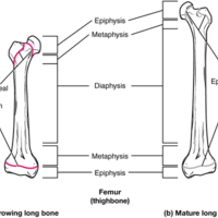
Progression from Epiphyseal Plate to Epiphyseal Line.jpg
As a bone matures, the epiphyseal plate progresses to an epiphyseal line. (a) Epiphyseal plates are visible in a growing bone. (b) Epiphyseal lines are the remnants of epiphyseal plates in a mature bone.

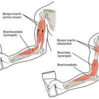
Prime Movers and Synergists.jpg
The biceps brachii flex the lower arm. The brachoradialis, in the forearm, and brachialis, located deep to the biceps in the upper arm, are both synergists that aid in this motion.

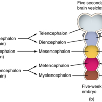
Primary and Secondary Vesicle Stages of Development.jpg
The embryonic brain develops complexity through enlargements of the neural tube called vesicles; (a) The primary vesicle stage has three regions, and (b) the secondary vesicle stage has five regions.

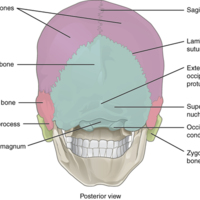
Posterior View of Skull.jpg
This view of the posterior skull shows attachment sites for muscles and joints that support the skull.

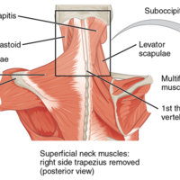
Posterior and Lateral Views of the Neck.jpg
The superficial and deep muscles of the neck are responsible for moving the head, cervical vertebrae, and scapulas.

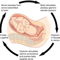
106_Pregnancy-Positive_Feedback.jpg
Normal childbirth is driven by a positive feedback loop. A positive feedback loop results in a change in the body’s status, rather than a return to homeostasis.

Photoreceptor.jpg
(a) All photoreceptors have inner segments containing the nucleus and other important organelles and outer segments with membrane arrays containing the photosensitive opsin molecules. Rod outer segments are long columnar shapes with stacks of…

Periosteum and Endosteum.jpg
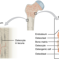
The periosteum forms the outer surface of bone, and the endosteum lines the medullary cavity.

Pelvis.jpg
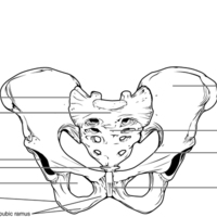
The pelvic girdle is formed by a single hip bone. The hip bone attaches the lower limb to the axial skeleton through its articulation with the sacrum. The right and left hip bones, plus the sacrum and the coccyx, together form the pelvis.

Pectoral Girdle.jpg
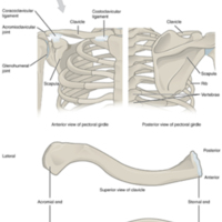
The pectoral girdle consists of the clavicle and the scapula, which serve to attach the upper limb to the sternum of the axial skeleton.

Pathways in Calcium Homeostasis.jpg
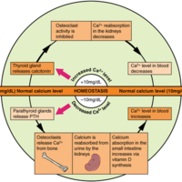
The body regulates calcium homeostasis with two pathways; one is signaled to turn on when blood calcium levels drop below normal and one is the pathway that is signaled to turn on when blood calcium levels are elevated.

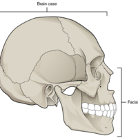
Parts of the Skull.jpg
The skull consists of the rounded brain case that houses the brain and the facial bones that form the upper and lower jaws, nose, orbits, and other facial structures.

Parts of a Typical Vertebra.jpg
A typical vertebra consists of a body and a vertebral arch. The arch is formed by the paired pedicles and paired laminae. Arising from the vertebral arch are the transverse, spinous, superior articular, and inferior articular processes. The vertebral…

Parts of a Neuron.jpg
The major parts of the neuron are labeled on a multipolar neuron from the CNS.

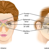
Paranasal Sinuses.jpg
The paranasal sinuses are hollow, air-filled spaces named for the skull bone that each occupies. The most anterior is the frontal sinus, located in the frontal bone above the eyebrows. The largest are the maxillary sinuses, located in the right and…

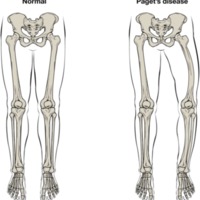
Paget's Disease.png
Normal leg bones are relatively straight, but those affected by Paget’s disease are porous and curved.

Overview of the Muscular System.jpg
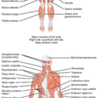
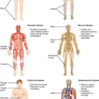
On the anterior and posterior views of the muscular system above, superficial muscles (those at the surface) are shown on the right side of the body while deep muscles (those underneath the superficial muscles) are shown on the left half of the body.…

Other Neuron Classifications.jpg
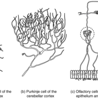
Three examples of neurons that are classified on the basis of other criteria. (a) The pyramidal cell is a multipolar cell with a cell body that is shaped something like a pyramid. (b) The Purkinje cell in the cerebellum was named after the scientist…

Osteoporosis.jpg
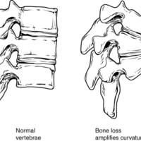
Osteoporosis is an age-related disorder that causes the gradual loss of bone density and strength. When the thoracic vertebrae are affected, there can be a gradual collapse of the vertebrae. This results in kyphosis, an excessive curvature of the…

Osteoarthritis.jpg
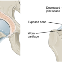
Osteoarthritis of a synovial joint results from aging or prolonged joint wear and tear. These cause erosion and loss of the articular cartilage covering the surfaces of the bones, resulting in inflammation that causes joint stiffness and pain.

Optic Nerve Versus Optic Tract.jpg
This drawing of the connections of the eye to the brain shows the optic nerve extending from the eye to the chiasm, where the structure continues as the optic tract. The same axons extend from the eye to the brain through these two bundles of fibers,…

Newborn Skull.jpg
The bones of the newborn skull are not fully ossified and are separated by large areas called fontanelles, which are filled with fibrous connective tissue. The fontanelles allow for continued growth of the skull after birth. At the time of birth, the…

Neuron Classification by Shape.jpg
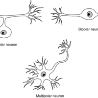
Unipolar cells have one process that includes both the axon and dendrite. Bipolar cells have two processes, the axon and a dendrite. Multipolar cells have more than two processes, the axon and two or more dendrites.

Nervous Tissue.jpg
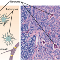
Nervous tissue is made up of neurons and neuroglia. The cells of nervous tissue are specialized to transmit and receive impulses

Nerve Structure.jpg
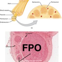
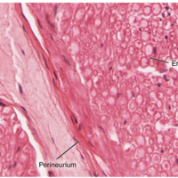
The structure of a nerve is organized by the layers of connective tissue on the outside, around each fascicle, and surrounding the individual nerve fibers (tissue source: simian).

Nerve Plexuses of the Body.jpg
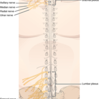
There are four main nerve plexuses in the human body. The cervical plexus supplies nerves to the posterior head and neck, as well as to the diaphragm. The brachial plexus supplies nerves to the arm. The lumbar plexus supplies nerves to the anterior…

Nasal Septum.jpg
The nasal septum is formed by the perpendicular plate of the ethmoid bone and the vomer bone. The septal cartilage fills the gap between these bones and extends into the nose.

Nails.jpg
The nail is an accessory structure of the integumentary system.

Muscles That Position the Pectoral Girdle.jpg
The muscles that stabilize the pectoral girdle make it a steady base on which other muscles can move the arm. Note that the pectoralis major and deltoid, which move the humerus, are cut here to show the deeper positioning muscles.

Muscles That Move the Tongue.jpg
Tongue muscles can be extrinsic or intrinsic. Extrinsic tongue muscles insert into the tongue from outside origins, and the intrinsic tongue muscles insert into the tongue from origins within it. The extrinsic muscles move the whole tongue in…

Muscles That Move the Lower Jaw.jpg
The muscles that move the lower jaw are typically located within the cheek and originate from processes in the skull. This provides the jaw muscles with the large amount of leverage needed for chewing.

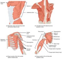
Muscles That Move the Humerus.jpg
(a, c) The muscles that move the humerus anteriorly are generally located on the anterior side of the body and originate from the sternum (e.g., pectoralis major) or the anterior side of the scapula (e.g., subscapularis). (b) The muscles that move…

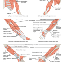
Muscles That Move the Forearm.jpg
The muscles originating in the upper arm flex, extend, pronate, and supinate the forearm. The muscles originating in the forearm move the wrists, hands, and fingers.

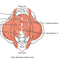
Muscles of the Pelvic Floor.jpg
The pelvic floor muscles support the pelvic organs, resist intra-abdominal pressure, and work as sphincters for the urethra, rectum, and vagina.

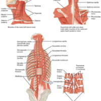
Muscles of the Neck and Back.jpg
The large, complex muscles of the neck and back move the head, shoulders, and vertebral column.

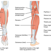
Muscles of the Lower Leg.jpg
The muscles of the anterior compartment of the lower leg are generally responsible for dorsiflexion, and the muscles of the posterior compartment of the lower leg are generally responsible for plantar flexion. The lateral and medial muscles in both…

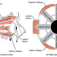
Muscles of the Eyes.jpg
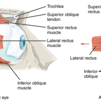
(a) The extrinsic eye muscles originate outside of the eye on the skull. (b) Each muscle inserts onto the eyeball.

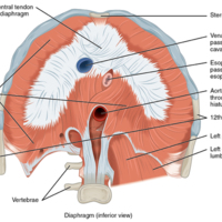
Muscles of the Diaphragm.jpg
The diaphragm separates the thoracic and abdominal cavities.

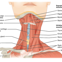
Muscles of the Anterior Neck.jpg
The anterior muscles of the neck facilitate swallowing and speech. The suprahyoid muscles originate from above the hyoid bone in the chin region. The infrahyoid muscles originate below the hyoid bone in the lower neck.

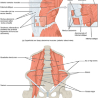
Muscles of the Abdomen.jpg
(a) The anterior abdominal muscles include the medially located rectus abdominis, which is covered by a sheet of connective tissue called the rectus sheath. On the flanks of the body, medial to the rectus abdominis, the abdominal wall is composed of…

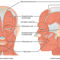
Muscles of Facial Expression.jpg
Many of the muscles of facial expression insert into the skin surrounding the eyelids, nose and mouth, producing facial expressions by moving the skin rather than bones.

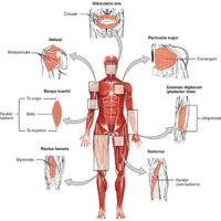
Muscle Shapes and Fiber Alignment.jpg
The skeletal muscles of the body typically come in seven different general shapes.

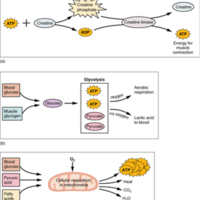
Muscle Metabolism.jpg
(a) Some ATP is stored in a resting muscle. As contraction starts, it is used up in seconds. More ATP is generated from creatine phosphate for about 15 seconds. (b) Each glucose molecule produces two ATP and two molecules of pyruvic acid, which can…

Muscle Fiber.jpg
A skeletal muscle fiber is surrounded by a plasma membrane called the sarcolemma, which contains sarcoplasm, the cytoplasm of muscle cells. A muscle fiber is composed of many fibrils, which give the cell its striated appearance.

Muscle Contraction.jpg
The dense bodies and intermediate filaments are networked through the sarcoplasm, which cause the muscle fiber to contract.

Multinucleate Muscle Cell.jpg
Unlike cardiac muscle cells and smooth muscle cells, which have a single nucleus, a skeletal muscle cell contains many nuclei, and is referred to as “multinucleated.” These muscle cells are long and fibrous (often referred to as muscle fibers).…

Multiaxial Joint.jpg
A multiaxial joint, such as the hip joint, allows for three types of movement: anterior-posterior, medial-lateral, and rotational.

Movements of the Body, Part 2.jpg
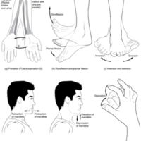
(g) Supination of the forearm turns the hand to the palm forward position in which the radius and ulna are parallel, while forearm pronation turns the hand to the palm backward position in which the radius crosses over the ulna to form an "X." (h)…

Movements of the Body, Part 1.jpg
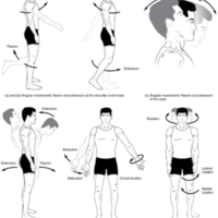
Synovial joints give the body many ways in which to move. (a)–(b) Flexion and extension motions are in the sagittal (anterior–posterior) plane of motion. These movements take place at the shoulder, hip, elbow, knee, wrist, metacarpophalangeal,…

Motor Units.jpg
A series of axon-like swelling, called varicosities or “boutons,” from autonomic neurons form motor units through the smooth muscle.

Motor End-Plate and Innervation.jpg
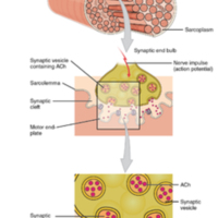
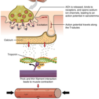
At the NMJ, the axon terminal releases ACh. The motor end-plate is the location of the ACh-receptors in the muscle fiber sarcolemma. When ACh molecules are released, they diffuse across a minute space called the synaptic cleft and bind to the…

Moles.jpg
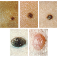
Moles range from benign accumulations of melanocytes to melanomas. These structures populate the landscape of our skin.

Molecular Structure of DNA.jpg
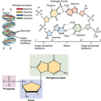
The DNA double helix is composed of two complementary strands. The strands are bonded together via their nitrogenous base pairs using hydrogen bonds.

Modes of Glandular Secretion.jpg
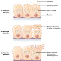
(a) In merocrine secretion, the cell remains intact. (b) In apocrine secretion, the apical portion of the cell is released, as well. (c) In holocrine secretion, the cell is destroyed as it releases its product and the cell itself becomes part of the…

Mitochondrion.jpg
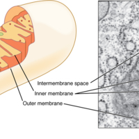
The mitochondria are the energy-conversion factories of the cell. (a) A mitochondrion is composed of two separate lipid bilayer membranes. Along the inner membrane are various molecules that work together to produce ATP, the cell’s major energy…

https://i.ytimg.com/vi/4g-RLu6afrY/default.jpg
The anatomy shown in this video is based off of the UBC MEDD 411 gross anatomy checklist.

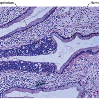
Micrograph of Cervical Tissue.jpg
This figure is a view of the regular architecture of normal tissue contrasted with the irregular arrangement of cancerous cells.

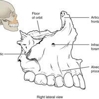
Maxillary Bone.jpg
The maxillary bone forms the upper jaw and supports the upper teeth. Each maxilla also forms the lateral floor of each orbit and the majority of the hard palate.

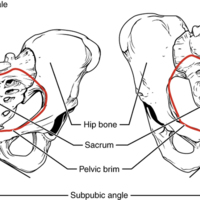
Male and Female Pelvis.jpg
The female pelvis is adapted for childbirth and is broader, with a larger subpubic angle, a rounder pelvic brim, and a wider and more shallow lesser pelvic cavity than the male pelvis.

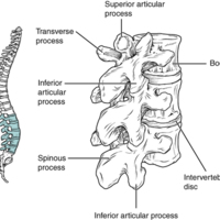
Lumbar Vertebrae.jpg
Lumbar vertebrae are characterized by having a large, thick body and a short, rounded spinous process.

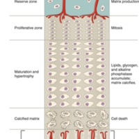
Longitudinal Bone Growth.jpg
The epiphyseal plate is responsible for longitudinal bone growth.

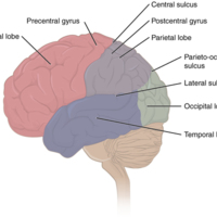
Lobes of the Cerebral Cortex.jpg
The cerebral cortex is divided into four lobes. Extensive folding increases the surface area available for cerebral functions.

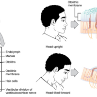
Linear Acceleration Coding by Maculae.jpg
The maculae are specialized for sensing linear acceleration, such as when gravity acts on the tilting head, or if the head starts moving in a straight line. The difference in inertia between the hair cell stereocilia and the otolithic membrane in…

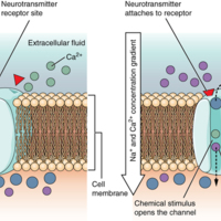
Ligand-Gated Channels.jpg
When the ligand, in this case the neurotransmitter acetylcholine, binds to a specific location on the extracellular surface of the channel protein, the pore opens to allow select ions through. The ions, in this case, are cations of sodium, calcium,…

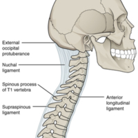
Ligaments of Vertebral Column.jpg
The anterior longitudinal ligament runs the length of the vertebral column, uniting the anterior sides of the vertebral bodies. The supraspinous ligament connects the spinous processes of the thoracic and lumbar vertebrae. In the posterior neck, the…

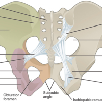
Ligaments of the Pelvis.jpg
The posterior sacroiliac ligament supports the sacroiliac joint. The sacrospinous ligament spans the sacrum to the ischial spine, and the sacrotuberous ligament spans the sacrum to the ischial tuberosity. The sacrospinous and sacrotuberous ligaments…

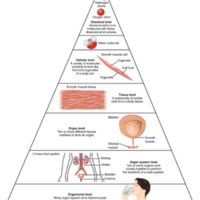
101_Levels_of_Org_in_Body.jpg
Before you begin to study the different structures and functions of the human body, it is helpful to consider its basic architecture; that is, how its smallest parts are assembled into larger structures. It is convenient to consider the structures of…

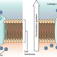
Leakage Channels.jpg
In certain situations, ions need to move across the membrane randomly. The particular electrical properties of certain cells are modified by the presence of this type of channel.

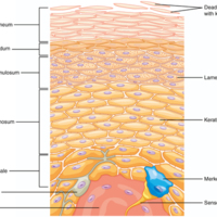
Layers of the Epidermis.jpg
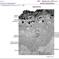
The epidermis of thick skin has five layers: stratum basale, stratum spinosum, stratum granulosum, stratum lucidum, and stratum corneum.

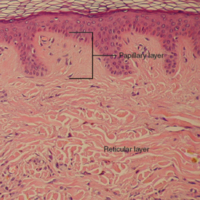
Layers of the Dermis.jpg
This stained slide shows the two components of the dermis—the papillary layer and the reticular layer. Both are made of connective tissue with fibers of collagen extending from one to the other, making the border between the two somewhat indistinct.…

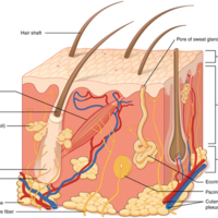
Layers of Skin.jpg
The skin is composed of two main layers: the epidermis, made of closely packed epithelial cells, and the dermis, made of dense, irregular connective tissue that houses blood vessels, hair follicles, sweat glands, and other structures. Beneath the…

Lateral Wall of Nasal Cavity.jpg
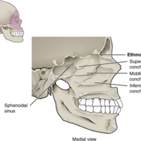
The three nasal conchae are curved bones that project from the lateral walls of the nasal cavity. The superior nasal concha and middle nasal concha are parts of the ethmoid bone. The inferior nasal concha is an independent bone of the sku

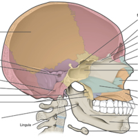
Lateral View of Skull.jpg
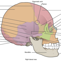
The lateral skull shows the large rounded brain case, zygomatic arch, and the upper and lower jaws. The zygomatic arch is formed jointly by the zygomatic process of the temporal bone and the temporal process of the zygomatic bone. The shallow space…

Knee Joint.jpg
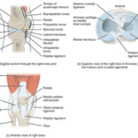
(a) The knee joint is the largest joint of the body. (b)–(c) It is supported by the tibial and fibular collateral ligaments located on the sides of the knee outside of the articular capsule, and the anterior and posterior cruciate ligaments found…

Knee Injury.jpg
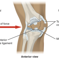
A strong blow to the lateral side of the extended knee will cause three injuries, in sequence: tearing of the tibial collateral ligament, damage to the medial meniscus, and rupture of the anterior cruciate ligament.

Isolated Mandible.jpg
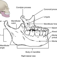
The mandible is the only moveable bone of the skull.

1. Introduction to Human Osteology.pdf
Physical anthropologists study human biological variation in the past and present. They are not only interested in the physical aspect of the body but also how biology, culture and environment interact to produce variation. Part of this variation…

Intrinsic Muscles of the Hand.jpg
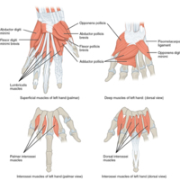
The intrinsic muscles of the hand both originate and insert within the hand. These muscles provide the fine motor control of the fingers by flexing, extending, abducting, and adducting the more distal finger and thumb segments.

Intrinsic Muscles of the Foot.jpg
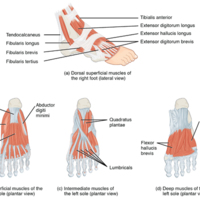
The muscles along the dorsal side of the foot (a) generally extend the toes while the muscles of the plantar side of the foot (b, c, d) generally flex the toes. The plantar muscles exist in three layers, providing the foot the strength to…

Intramembranous Ossification.jpg
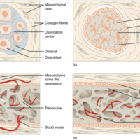
Intramembranous ossification follows four steps. (a) Mesenchymal cells group into clusters, and ossification centers form. (b) Secreted osteoid traps osteoblasts, which then become osteocytes. (c) Trabecular matrix and periosteum form. (d) Compact…

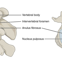
Intervertebral Disc.jpg
The bodies of adjacent vertebrae are separated and united by an intervertebral disc, which provides padding and allows for movements between adjacent vertebrae. The disc consists of a fibrous outer layer called the anulus fibrosus and a gel-like…

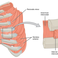
Intercostal Muscles.jpg
The external intercostals are located laterally on the sides of the body. The internal intercostals are located medially near the sternum. The innermost intercostals are located deep to both the internal and external intercostals.

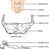
Hyoid Bone.jpg
The hyoid bone is located in the upper neck and does not join with any other bone. It provides attachments for muscles that act on the tongue, larynx, and pharynx.

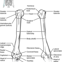
Humerus and Elbow Joint.jpg
The humerus is the single bone of the upper arm region. It articulates with the radius and ulna bones of the forearm to form the elbow joint.

Human-Anatomy-Lab-Manual-1535056958._print.pdf
This is a lab manual for a college-level human anatomy course. Mastery of anatomy requires a fair amount of memorization and recall skills. The activities in this manual encourage students to engage with new vocabulary in many ways, including…

Human Anatomy and Physiology Preparatory Course.pdf
The overall purpose of this preparatory course textbook is to help students familiarize with some terms and some basic concepts they will find later in the Human Anatomy and Physiology I course. The organization and functioning of the human organism…

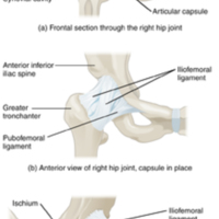
Hip Joint.jpg
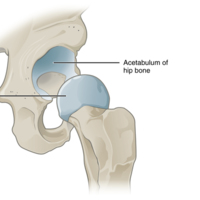
(a) The ball-and-socket joint of the hip is a multiaxial joint that provides both stability and a wide range of motion. (b–c) When standing, the supporting ligaments are tight, pulling the head of the femur into the acetabulum.

Hip and Thigh Muscles.jpg
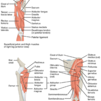
The large and powerful muscles of the hip that move the femur generally originate on the pelvic girdle and insert into the femur. The muscles that move the lower leg typically originate on the femur and insert into the bones of the knee joint. The…

Herniated Intervertebral Disc.jpg
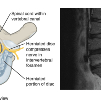
Weakening of the anulus fibrosus can result in herniation (protrusion) of the nucleus pulposus and compression of a spinal nerve, resulting in pain and/or muscle weakness in the body regions supplied by that nerve.

Hematopoiesis.jpg
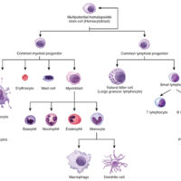
The process of hematopoiesis involves the differentiation of multipotent cells into blood and immune cells. The multipotent hematopoietic stem cells give rise to many different cell types, including the cells of the immune system and red blood cells.

Hand During Gripping.jpg
During tight gripping—compare (b) to (a)—the fourth and, particularly, the fifth metatarsal bones are pulled anteriorly. This increases the contact between the object and the medial side of the hand, thus improving the firmness of the grip.

Hair Cell.jpg
The hair cell is a mechanoreceptor with an array of stereocilia emerging from its apical surface. The stereocilia are tethered together by proteins that open ion channels when the array is bent toward the tallest member of their array, and closed…

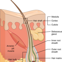
Hair.jpg
Hair follicles originate in the epidermis and have many different parts.

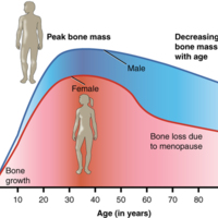
Graph Showing Relationship Between Age and Bone Mass.jpg
Bone density peaks at about 30 years of age. Women lose bone mass more rapidly than men.

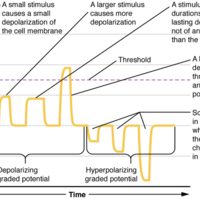
Graded Potentials.jpg
Graded potentials are temporary changes in the membrane voltage, the characteristics of which depend on the size of the stimulus. Some types of stimuli cause depolarization of the membrane, whereas others cause hyperpolarization. It depends on the…

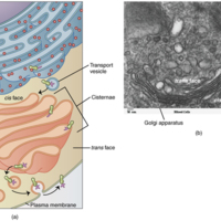
Golgi Apparatus.jpg
(a) The Golgi apparatus manipulates products from the rough ER, and also produces new organelles called lysosomes. Proteins and other products of the ER are sent to the Golgi apparatus, which organizes, modifies, packages, and tags them. Some of…

Goblet Cell.jpg
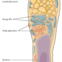
(a) In the lining of the small intestine, columnar epithelium cells are interspersed with goblet cells. (b) The arrows in this micrograph point to the mucous-secreting goblet cells.

Glial Cells of the PNS.jpg
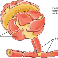
The PNS has satellite cells and Schwann cells.

Glial Cells of the CNS.jpg
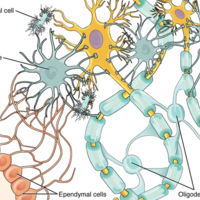
The CNS has astrocytes, oligodendrocytes, microglia, and ependymal cells that support the neurons of the CNS in several ways.

Glenohumeral Joint.jpg
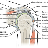
The glenohumeral (shoulder) joint is a ball-and-socket joint that provides the widest range of motions. It has a loose articular capsule and is supported by ligaments and the rotator cuff muscles.

Manz-HumanAnatomy-Cover-r-350x453.png
This laboratory manual is meant to be used in a lab course that accompanies a college-level introductory human anatomy course. It covers the language of anatomy, cells and tissues, and the organ systems that comprise the human body. Multiple open…

A-and-P-cover_final-350x559.png
ght in Australia, aiming to provide students with an increased access to free, high-quality learning materials.

The material in this textbook is largely based on OpenStax’s Anatomy & Physiology textbook, however, has been modified for Australian…
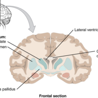
Frontal Section of Cerebral Cortex and Basal Nuclei.jpg
The major components of the basal nuclei, shown in a frontal section of the brain, are the caudate (just lateral to the lateral ventricle), the putamen (inferior to the caudate and separated by the large white-matter structure called the internal…

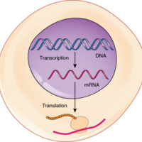
From DNA to Protein Transcription through Translation.jpg
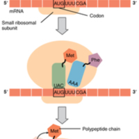
Transcription within the cell nucleus produces an mRNA molecule, which is modified and then sent into the cytoplasm for translation. The transcript is decoded into a protein with the help of a ribosome and tRNA molecules.

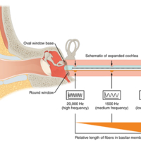
Frequency Coding in the Cochlea.jpg
The standing sound wave generated in the cochlea by the movement of the oval window deflects the basilar membrane on the basis of the frequency of sound. Therefore, hair cells at the base of the cochlea are activated only by high frequencies, whereas…

Fractures of the Humerus and Radius.jpg
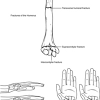
Falls or direct blows can result in fractures of the surgical neck or shaft of the humerus. Falls onto the elbow can fracture the distal humerus. A Colles fracture of the distal radius is the most common forearm fracture.

Four Types of Tissue Body.jpg
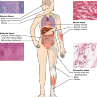
The four types of tissues are exemplified in nervous tissue, stratified squamous epithelial tissue, cardiac muscle tissue, and connective tissue in small intestine. Clockwise from nervous tissue

Fluorescence-stained Cell Undergoing Mitosis.jpg
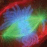
A lung cell from a newt, commonly studied for its similarity to human lung cells, is stained with fluorescent dyes. The green stain reveals mitotic spindles, red is the cell membrane and part of the cytoplasm, and the structures that appear light…

Fibrous Joints.jpg
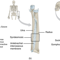
Fibrous joints form strong connections between bones. (a) Sutures join most bones of the skull. (b) An interosseous membrane forms a syndesmosis between the radius and ulna bones of the forearm. (c) A gomphosis is a specialized fibrous joint that…

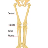
Femur and Patella.jpg
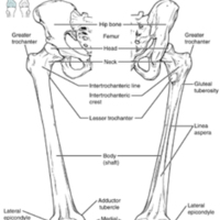
The femur is the single bone of the thigh region. It articulates superiorly with the hip bone at the hip joint, and inferiorly with the tibia at the knee joint. The patella only articulates with the distal end of the femur.

Fatty Acid Shapes.jpg
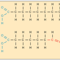
The level of saturation of a fatty acid affects its shape. (a) Saturated fatty acid chains are straight. (b) Unsaturated fatty acid chains are kinked.

Tags: , ,

Facilitated Diffusion.jpg
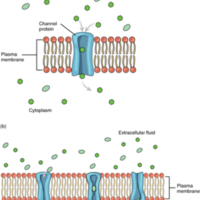
(a) Facilitated diffusion of substances crossing the cell (plasma) membrane takes place with the help of proteins such as channel proteins and carrier proteins. Channel proteins are less selective than carrier proteins, and usually mildly…

Extraocular Muscles.jpg
The extraocular muscles move the eye within the orbit.

External and Internal Views of Base of Skull.jpg
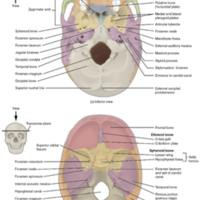
(a) The hard palate is formed anteriorly by the palatine processes of the maxilla bones and posteriorly by the horizontal plate of the palatine bones. (b) The complex floor of the cranial cavity is formed by the frontal, ethmoid, sphenoid, temporal,…

Exocytosis.jpg
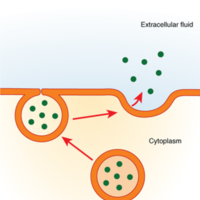
Exocytosis is much like endocytosis in reverse. Material destined for export is packaged into a vesicle inside the cell. The membrane of the vesicle fuses with the cell membrane, and the contents are released into the extracellular space.

excretory_pd_PearsonScottForesman.png
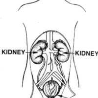
After food goes through the digestive system, the parts that are not digested need to be gotten rid of. That is the job of the excretory system.

Unabsorbed food goes to the large intestine. The liver also filters out solid particles of waste from…

Ethmoid Bone.jpg
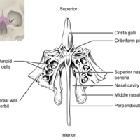
The unpaired ethmoid bone is located at the midline within the central skull. It has an upward projection, the crista galli, and a downward projection, the perpendicular plate, which forms the upper nasal septum. The cribriform plates form both the…

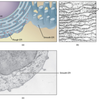
Endoplasmic Reticulum (ER).jpg
(a) The ER is a winding network of thin membranous sacs found in close association with the cell nucleus. The smooth and rough endoplasmic reticula are very different in appearance and function (source: mouse tissue). (b) Rough ER is studded with…

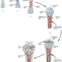
Endochondral Ossification.jpg
Endochondral ossification follows five steps. (a) Mesenchymal cells differentiate into chondrocytes. (b) The cartilage model of the future bony skeleton and the perichondrium form. (c) Capillaries penetrate cartilage. Perichondrium transforms into…

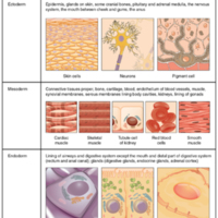
Embryonic Origin of Tissues.jpg
The zygote, or fertilized egg, is a single cell formed by the fusion of an egg and sperm. After fertilization the zygote gives rise to rapid mitotic cycles, generating many cells to form the embryo. The first embryonic cells generated have the…

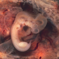
Embryo at Seven Weeks.jpg
Limb buds are visible in an embryo at the end of the seventh week of development (embryo derived from an ectopic pregnancy)

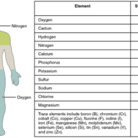
Elements of the Human Body.jpg
The main elements that compose the human body are shown from most abundant to least abundant.

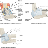
Elbow Joint.jpg
(a) The elbow is a hinge joint that allows only for flexion and extension of the forearm. (b) It is supported by the ulnar and radial collateral ligaments. (c) The annular ligament supports the head of the radius at the proximal radioulnar joint, the…

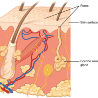
Eccrine Gland.jpg
Eccrine glands are coiled glands in the dermis that release sweat that is mostly water.

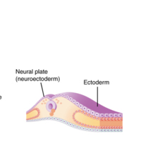
Early Embryonic Development of Nervous System.jpg
The neuroectoderm begins to fold inward to form the neural groove. As the two sides of the neural groove converge, they form the neural tube, which lies beneath the ectoderm. The anterior end of the neural tube will develop into the brain, and the…

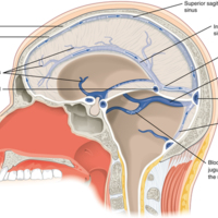
Dural Sinuses and Veins.jpg
Blood drains from the brain through a series of sinuses that connect to the jugular veins.

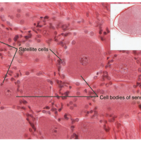
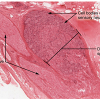
Dorsal Root Ganglion.jpg
The cell bodies of sensory neurons, which are unipolar neurons by shape, are seen in this photomicrograph. Also, the fibrous region is composed of the axons of these neurons that are passing through the ganglion to be part of the dorsal nerve root…

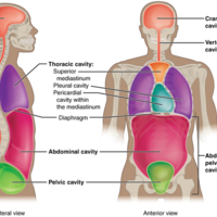
110_Dorsal_Ventral_Body_Cavities.jpg
The posterior (dorsal) and anterior (ventral) cavities are each subdivided into smaller cavities. In the posterior (dorsal) cavity, the cranial cavity houses the brain, and the spinal cavity (or vertebral cavity) encloses the spinal cord. Just as the…

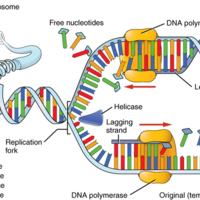
DNA Replication.jpg
DNA replication faithfully duplicates the entire genome of the cell. During DNA replication, a number of different enzymes work together to pull apart the two strands so each strand can be used as a template to synthesize new complementary strands.…

DNA Macrostructure.jpg
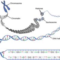
Strands of DNA are wrapped around supporting histones. These proteins are increasingly bundled and condensed into chromatin, which is packed tightly into chromosomes when the cell is ready to divide.

digesive_sys_PD_PearsonScottForesman.jpg
Our bodies need food to live and grow. The digestive system takes food and carries it to all the parts of the body.

The beginning of the digestive system is the mouth and teeth. Food that we eat has to be broken down into nutrients that cells in…

Diagram of Spongy Bone.jpg
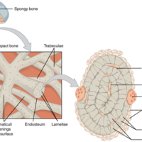
Spongy bone is composed of trabeculae that contain the osteocytes. Red marrow fills the spaces in some bones.

Diagram of Compact Bone.jpg
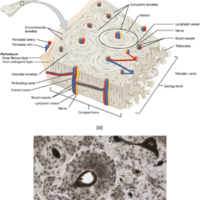
(a) This cross-sectional view of compact bone shows the basic structural unit, the osteon. (b) In this micrograph of the osteon, you can clearly see the concentric lamellae and central canals.

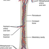
Diagram of Blood and Nerve Supply to Bone.jpg
Blood vessels and nerves enter the bone through the nutrient foramen.

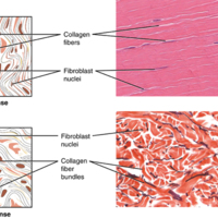
Dense Connective Tissue.jpg
(a) Dense regular connective tissue consists of collagenous fibers packed into parallel bundles. (b) Dense irregular connective tissue consists of collagenous fibers interwoven into a mesh-like network. From top,

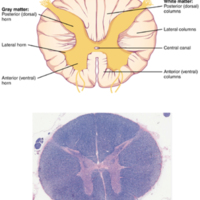
Cross-section of Spinal Cord.jpg
The cross-section of a thoracic spinal cord segment shows the posterior, anterior, and lateral horns of gray matter, as well as the posterior, anterior, and lateral columns of white matter

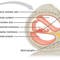
Cross Section of the Cochlea.jpg
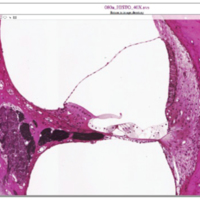
The three major spaces within the cochlea are highlighted. The scala tympani and scala vestibuli lie on either side of the cochlear duct. The organ of Corti, containing the mechanoreceptor hair cells, is adjacent to the scala tympani, where it sits…

Cranial Fossae.jpg
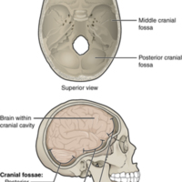
The bones of the brain case surround and protect the brain, which occupies the cranial cavity. The base of the brain case, which forms the floor of cranial cavity, is subdivided into the shallow anterior cranial fossa, the middle cranial fossa, and…

Corticospinal Tract.jpg
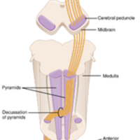
The major descending tract that controls skeletal muscle movements is the corticospinal tract. It is composed of two neurons, the upper motor neuron and the lower motor neuron. The upper motor neuron has its cell body in the primary motor cortex of…

Contraction of a Muscle Fiber.jpg
A cross-bridge forms between actin and the myosin heads triggering contraction. As long as Ca++ ions remain in the sarcoplasm to bind to troponin, and as long as ATP is available, the muscle fiber will continue to shorten.

Connective Tissue Proper.jpg
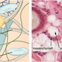
Fibroblasts produce this fibrous tissue. Connective tissue proper includes the fixed cells fibrocytes, adipocytes, and mesenchymal cells.

Connections of Parasympathetic Division of the Autonomic Nervous System.jpg
Neurons from brain-stem nuclei, or from the lateral horn of the sacral spinal cord, project to terminal ganglia near or within the various organs of the body. Axons from these ganglionic neurons then project the short distance to those target…

Comparison of Somatic and Visceral Reflexes.jpg
The afferent inputs to somatic and visceral reflexes are essentially the same, whereas the efferent branches are different. Somatic reflexes, for instance, involve a direct connection from the ventral horn of the spinal cord to the skeletal muscle.…

Comparison of Color Sensitivity of Photopigments.jpg
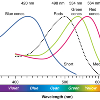
Comparing the peak sensitivity and absorbance spectra of the four photopigments suggests that they are most sensitive to particular wavelengths.

Cochlea and Organ of Corti.jpg
a given region of the basilar membrane will only move if the incoming sound is at a specific frequency. Because the tectorial membrane only moves where the basilar membrane moves, the hair cells in this region will also only respond to sounds of this…

Clubfoot.jpg
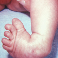
Clubfoot
This photograph shows a baby with a clubfoot.Clubfoot is a common deformity of the ankle and foot that is present at birth. Most cases are corrected without surgery, and affected individuals will grow up to lead normal, active lives.…

Close-Up of Nerve Trunk.jpg
Zoom in on this slide of a nerve trunk to examine the endoneurium, perineurium, and epineurium in greater detail (tissue source: simian).

Classifications of Bones.jpg
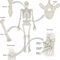
Bones are classified according to their shape.

Human Anatomy_Circulatory System.jpg
The circulatory system includes the heart, blood, and a huge network of blood vessels that carry blood all over the body. The job of the circulatory system is to deliver oxygen to cells all over the body and then to carry out waste product like…

Circle of Willis.jpg
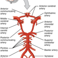
The blood supply to the brain enters through the internal carotid arteries and the vertebral arteries, eventually giving rise to the circle of Willis.

Human Anatomy6.jpg
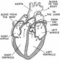
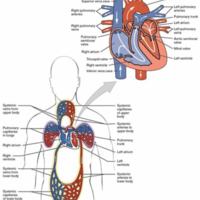
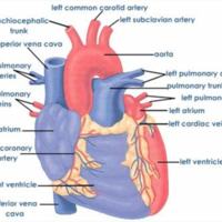
Dual System of the Human Blood Circulation. Blood flows from the right atrium to the right ventricle, where it is pumped into the pulmonary circuit. The blood in the pulmonary artery branches is low in oxygen but relatively high in carbon dioxide.…

Cervical Vertebrae.jpg
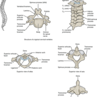
A typical cervical vertebra has a small body, a bifid spinous process, transverse processes that have a transverse foramen and are curved for spinal nerve passage. The atlas (C1 vertebra) does not have a body or spinous process. It consists of an…

Cerebrospinal Fluid Circulation.jpg
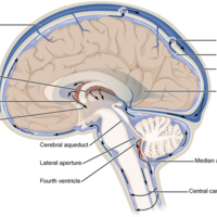
The choroid plexus in the four ventricles produce CSF, which is circulated through the ventricular system and then enters the subarachnoid space through the median and lateral apertures. The CSF is then reabsorbed into the blood at the arachnoid…

Central and Peripheral Nervous System.jpg
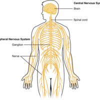
The structures of the PNS are referred to as ganglia and nerves, which can be seen as distinct structures. The equivalent structures in the CNS are not obvious from this overall perspective and are best examined in prepared tissue under the…

Cells of the Epidermis.jpg
The cells in the different layers of the epidermis originate from basal cells located in the stratum basale, yet the cells of each layer are distinctively different.

Cells of Epithelial Tissue.jpg
Simple epithelial tissue is organized as a single layer of cells and stratified epithelial tissue is formed by several layers of cells.

Cell Membrane and Transmembrane Proteins.jpg
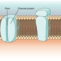
The cell membrane is composed of a phospholipid bilayer and has many transmembrane proteins, including different types of channel proteins that serve as ion channels.

Cell Membrane.jpg
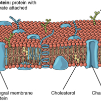
The cell membrane of the cell is a phospholipid bilayer containing many different molecular components, including proteins and cholesterol, some with carbohydrate groups attached.

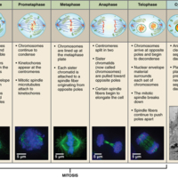
Cell Division Mitosis.jpg
The stages of cell division oversee the separation of identical genetic material into two new nuclei, followed by the division of the cytoplasm.

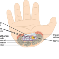
Carpal Tunnel.jpg
The carpal tunnel is the passageway by which nine muscle tendons and a major nerve enter the hand from the anterior forearm. The walls and floor of the carpal tunnel are formed by the U-shaped grouping of the carpal bones, and the roof is formed by…

Human Anatomy5.jpg
Cardio Exercises Below
1) Jog. You can do this outside on a treadmill or however you like.
2) Jump Rope routine 1
3) 10 Minute Jump Rope routine
4) Exercise Bikes
5) Sports Playing

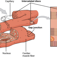
Cardiac Muscle.jpg
Intercalated discs are part of the cardiac muscle sarcolemma and they contain gap junctions and desmosomes.

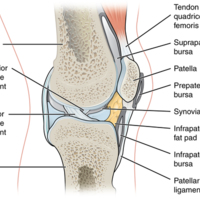
Bursae.jpg
Bursae are fluid-filled sacs that serve to prevent friction between skin, muscle, or tendon and an underlying bone. Three major bursae and a fat pad are part of the complex joint that unites the femur and tibia of the leg.

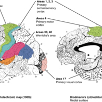
Brodmann's Areas of the Cerebral Cortex.jpg
Brodmann mapping of functionally distinct regions of the cortex was based on its cytoarchitecture at a microscopic level.

brehes-grammar-anatomy.pdf
Brehe’s Grammar Anatomy makes grammar accessible to general and specialist readers alike. This book provides an in-depth look at beginner grammar terms and concepts, providing clear examples with limited technical jargon. Whether for academic or…

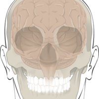
Bones Protect Brain.jpg
The cranium completely surrounds and protects the brain from non-traumatic injury.

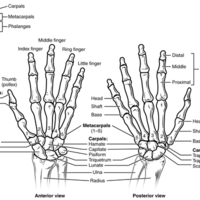
Bones of the Wrist and Hand.jpg
The eight carpal bones form the base of the hand. These are arranged into proximal and distal rows of four bones each. The metacarpal bones form the palm of the hand. The thumb and fingers consist of the phalanx bones.

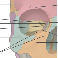
Bones of the Orbit.jpg
Seven skull bones contribute to the walls of the orbit. Opening into the posterior orbit from the cranial cavity are the optic canal and superior orbital fissure.

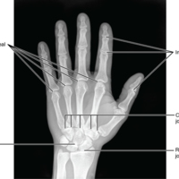
Bones of the Hand.jpg
This radiograph shows the position of the bones within the hand. Note the carpal bones that form the base of the hand

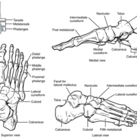
Bones of the Foot.jpg
The bones of the foot are divided into three groups. The posterior foot is formed by the seven tarsal bones. The mid-foot has the five metatarsal bones. The toes contain the phalanges.

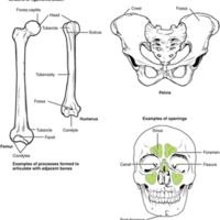
Bone Features.jpg
The surface features of bones depend on their function, location, attachment of ligaments and tendons, or the penetration of blood vessels and nerves.

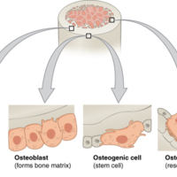
Bone Cells.jpg
Four types of cells are found within bone tissue. Osteogenic cells are undifferentiated and develop into osteoblasts. When osteoblasts get trapped within the calcified matrix, their structure and function changes, and they become osteocytes.…

Axial and Appendicular Skeleton.jpg
The axial skeleton supports the head, neck, back, and chest and thus forms the vertical axis of the body. It consists of the skull, vertebral column (including the sacrum and coccyx), and the thoracic cage, formed by the ribs and sternum. The…

Autonomic Varicosities.jpg
The connection between autonomic fibers and target effectors is not the same as the typical synapse, such as the neuromuscular junction. Instead of a synaptic end bulb, a neurotransmitter is released from swellings along the length of a fiber that…

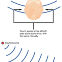
Auditory Brain Stem Mechanisms of Sound Localization.jpg
Localizing sound in the horizontal plane is achieved by processing in the medullary nuclei of the auditory system. Connections between neurons on either side are able to compare very slight differences in sound stimuli that arrive at either ear and…

Atrophy.png
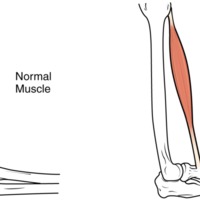
Muscle mass is reduced as muscles atrophy with disuse.

Atlantoaxial Joint.jpg
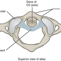
The atlantoaxial joint is a pivot type of joint between the dens portion of the axis (C2 vertebra) and the anterior arch of the atlas (C1 vertebra), with the dens held in place by a ligament.

Ascending Sensory Pathways of the Spinal Cord.jpg
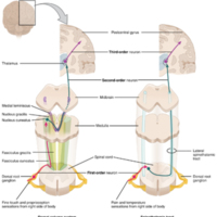
The dorsal column system and spinothalamic tract are the major ascending pathways that connect the periphery with the brain.

Anterior View of Skull.jpg
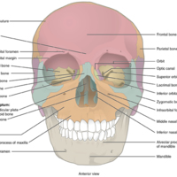
An anterior view of the skull shows the bones that form the forehead, orbits (eye sockets), nasal cavity, nasal septum, and upper and lower jaws.

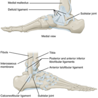
Ankle Joint.jpg
The talocrural (ankle) joint is a uniaxial hinge joint that only allows for dorsiflexion or plantar flexion of the foot. Movements at the subtalar joint, between the talus and calcaneus bones, combined with motions at other intertarsal joints,…

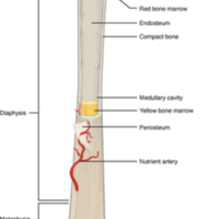
Anatomy of a Long Bone.jpg
A typical long bone shows the gross anatomical characteristics of bone.

Anatomy of a Flat Bone.jpg
This cross-section of a flat bone shows the spongy bone (diploë) lined on either side by a layer of compact bone.

Midterm-eye-3-350x623.jpg
Authors’ Institution: Norfolk State University, Norfolk, VA, USA , Funding: Virtual Library of Virginia (VIVA) Course Redesign Grant, 2020 – 2023

uh-oer-cover-anatomy-physiology-lab-prof-350x453.png
This manual is a guide for anatomy and physiology laboratory exercises. It includes dissection guidance with detailed images; instructions for physiology experiments including foundational content; and gross anatomy study guides for six body systems.…
2.pdf
Welcome to Human Anatomy and Physiology, an OpenStax College resource. We created this textbook with several goals
in mind: accessibility, customization, and student engagement—helping students reach high levels of academic scholarship.
Instructors…

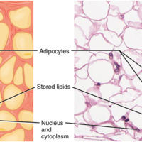
Adipose Tissue.jpg
This is a loose connective tissue that consists of fat cells with little extracellular matrix. It stores fat for energy and provides insulation.
Adenosine Triphosphate.jpg
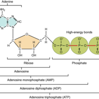
The nucleotide adenosine triphosphate (ATP), is composed of a ribose sugar, an adenine base, and three phosphate groups ([link]). ATP is classified as a high energy compound because the two covalent bonds linking its three phosphates store a…
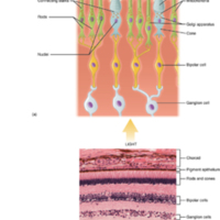
Acne.jpg
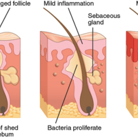
Acne is a result of over-productive sebaceous glands, which leads to formation of blackheads and inflammation of the skin.

Abnormal Curvatures of the Vertebral Column.jpg
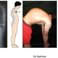
(a) Scoliosis is an abnormal lateral bending of the vertebral column. (b) An excessive curvature of the upper thoracic vertebral column is called kyphosis. (c) Lordosis is an excessive curvature in the lumbar region of the vertebral column.

A Homologous Pair of Chromosomes with their Attached Sister Chromatids.jpg
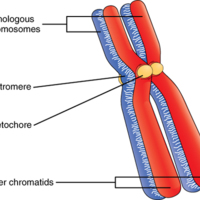
The red and blue colors correspond to a homologous pair of chromosomes. Each member of the pair was separately inherited from one parent. Each chromosome in the homologous pair is also bound to an identical sister chromatid, which is produced by DNA…
Output Formats

atom, dcmes-xml, json, omeka-xml, rss2