Browse Items (66 total)
- Tags: Medicine & Health
Sort by:
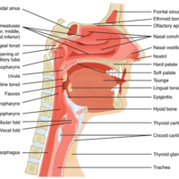
The Nose and its Adjacent Structures
Several bones that help form the walls of the nasal cavity have air-containing spaces called the paranasal sinuses, which serve to warm and humidify incoming air. Sinuses are lined with a mucosa. Each paranasal sinus is named for its associated bone:…
Comparison of Somatic and Visceral Reflexes
The afferent inputs to somatic and visceral reflexes are essentially the same, whereas the efferent branches are different. Somatic reflexes, for instance, involve a direct connection from the ventral horn of the spinal cord to the skeletal muscle.…
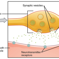
Autonomic Varicosities
The connection between autonomic fibers and target effectors is not the same as the typical synapse, such as the neuromuscular junction. Instead of a synaptic end bulb, a neurotransmitter is released from swellings along the length of a fiber that…
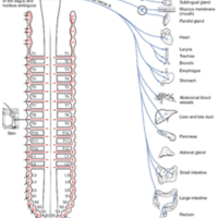
Connections of Parasympathetic Division of the Autonomic Nervous System
Neurons from brain-stem nuclei, or from the lateral horn of the sacral spinal cord, project to terminal ganglia near or within the various organs of the body. Axons from these ganglionic neurons then project the short distance to those target…
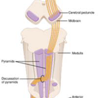
Corticospinal Tract
The major descending tract that controls skeletal muscle movements is the corticospinal tract. It is composed of two neurons, the upper motor neuron and the lower motor neuron. The upper motor neuron has its cell body in the primary motor cortex of…
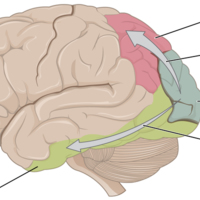
Ventral and Dorsal Visual Streams
From the primary visual cortex in the occipital lobe, visual processing continues in two streams—one into the temporal lobe and one into the parietal lobe.
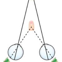
Retinal Disparity
Because of the interocular distance, which results in objects of different distances falling on different spots of the two retinae, the brain can extract depth perception from the two-dimensional information of the visual field.
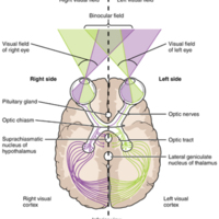
Topographic Mapping of the Retina onto the Visual Cortex
The visual field projects onto the retina through the lenses and falls on the retinae as an inverted, reversed image. The topography of this image is maintained as the visual information travels through the visual pathway to the cortex.
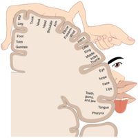
The Sensory Homunculus
A cartoon representation of the sensory homunculus arranged adjacent to the cortical region in which the processing takes place.
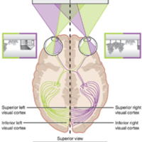
Segregation of Visual Field Information at the Optic Chiasm
Contralateral visual field information from the lateral retina projects to the ipsilateral brain, whereas ipsilateral visual field information has to decussate at the optic chiasm to reach the opposite side of the brain.
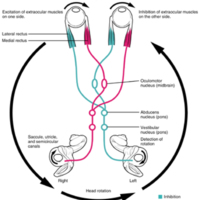
Vestibulo-ocular Reflex
Connections between the vestibular system and the cranial nerves controlling eye movement keep the eyes centered on a visual stimulus, even though the head is moving. During head movement, the eye muscles move the eyes in the opposite direction as…
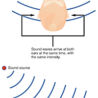
Auditory Brain Stem Mechanisms of Sound Localization
Localizing sound in the horizontal plane is achieved by processing in the medullary nuclei of the auditory system. Connections between neurons on either side are able to compare very slight differences in sound stimuli that arrive at either ear and…
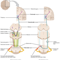
Ascending Sensory Pathways of the Spinal Cord
The dorsal column system and spinothalamic tract are the major ascending pathways that connect the periphery with the brain.
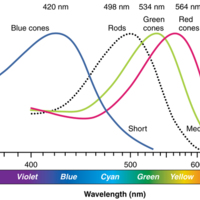
Comparison of Color Sensitivity of Photopigments
Comparing the peak sensitivity and absorbance spectra of the four photopigments suggests that they are most sensitive to particular wavelengths.
Retinal Isomers
The retinal molecule has two isomers, (a) one before a photon interacts with it and (b) one that is altered through photoisomerization.
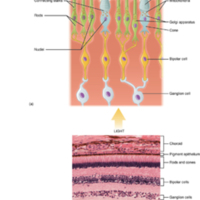
Photoreceptor
(a) All photoreceptors have inner segments containing the nucleus and other important organelles and outer segments with membrane arrays containing the photosensitive opsin molecules. Rod outer segments are long columnar shapes with stacks of…
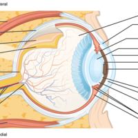
Structure of the Eye
The sphere of the eye can be divided into anterior and posterior chambers. The wall of the eye is composed of three layers: the fibrous tunic, vascular tunic, and neural tunic. Within the neural tunic is the retina, with three layers of cells and two…
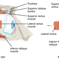
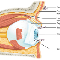
Extraocular Muscles
The extraocular muscles move the eye within the orbit.
The Eye in the Orbit
The eye is located within the orbit and surrounded by soft tissues that protect and support its function. The orbit is surrounded by cranial bones of the skull.
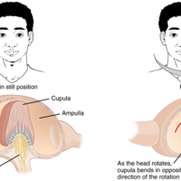
Rotational Coding by Semicircular Canals
Rotational movement of the head is encoded by the hair cells in the base of the semicircular canals. As one of the canals moves in an arc with the head, the internal fluid moves in the opposite direction, causing the cupula and stereocilia to bend.…
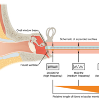
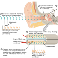
Frequency Coding in the Cochlea
The standing sound wave generated in the cochlea by the movement of the oval window deflects the basilar membrane on the basis of the frequency of sound. Therefore, hair cells at the base of the cochlea are activated only by high frequencies, whereas…
Cochlea and Organ of Corti
a given region of the basilar membrane will only move if the incoming sound is at a specific frequency. Because the tectorial membrane only moves where the basilar membrane moves, the hair cells in this region will also only respond to sounds of this…
Hair Cell
The hair cell is a mechanoreceptor with an array of stereocilia emerging from its apical surface. The stereocilia are tethered together by proteins that open ion channels when the array is bent toward the tallest member of their array, and closed…
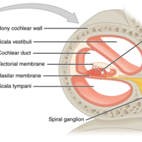
Cross Section of the Cochlea
The three major spaces within the cochlea are highlighted. The scala tympani and scala vestibuli lie on either side of the cochlear duct. The organ of Corti, containing the mechanoreceptor hair cells, is adjacent to the scala tympani, where it sits…
Transmission of Sound Waves to Cochlea
A sound wave causes the tympanic membrane to vibrate. This vibration is amplified as it moves across the malleus, incus, and stapes. The amplified vibration is picked up by the oval window causing pressure waves in the fluid of the scala vestibuli…
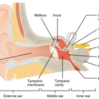
Structures of the Ear
The external ear contains the auricle, ear canal, and tympanic membrane. The middle ear contains the ossicles and is connected to the pharynx by the Eustachian tube. The inner ear contains the cochlea and vestibule, which are responsible for audition…
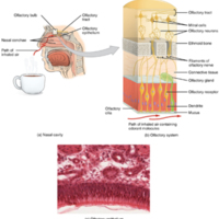
The Olfactory System
(a) The olfactory system begins in the peripheral structures of the nasal cavity. (b) The olfactory receptor neurons are within the olfactory epithelium. (c) Axons of the olfactory receptor neurons project through the cribriform plate of the ethmoid…
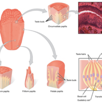
The Tongue
The tongue is covered with small bumps, called papillae, which contain taste buds that are sensitive to chemicals in ingested food or drink. Different types of papillae are found in different regions of the tongue. The taste buds contain specialized…
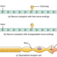
Receptor Classification by Cell Type
Receptor cell types can be classified on the basis of their structure. Sensory neurons can have either (a) free nerve endings or (b) encapsulated endings. Photoreceptors in the eyes, such as rod cells, are examples of (c) specialized receptor cells.…
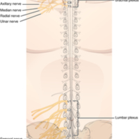
Nerve Plexuses of the Body
There are four main nerve plexuses in the human body. The cervical plexus supplies nerves to the posterior head and neck, as well as to the diaphragm. The brachial plexus supplies nerves to the arm. The lumbar plexus supplies nerves to the anterior…
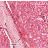
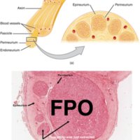
Close-Up of Nerve Trunk
Zoom in on this slide of a nerve trunk to examine the endoneurium, perineurium, and epineurium in greater detail (tissue source: simian).
Nerve Structure
The structure of a nerve is organized by the layers of connective tissue on the outside, around each fascicle, and surrounding the individual nerve fibers (tissue source: simian).
Spinal Cord and Root Ganglion
The slide includes both a cross-section of the lumbar spinal cord and a section of the dorsal root ganglion (see also [link]) (tissue source: canine).
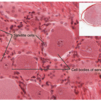
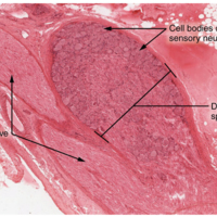
Dorsal Root Ganglion
The cell bodies of sensory neurons, which are unipolar neurons by shape, are seen in this photomicrograph. Also, the fibrous region is composed of the axons of these neurons that are passing through the ganglion to be part of the dorsal nerve root…
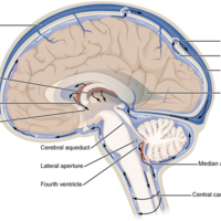
Cerebrospinal Fluid Circulation
The choroid plexus in the four ventricles produce CSF, which is circulated through the ventricular system and then enters the subarachnoid space through the median and lateral apertures. The CSF is then reabsorbed into the blood at the arachnoid…
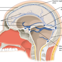
Dural Sinuses and Veins
Blood drains from the brain through a series of sinuses that connect to the jugular veins.
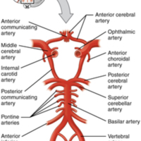
Circle of Willis
The blood supply to the brain enters through the internal carotid arteries and the vertebral arteries, eventually giving rise to the circle of Willis.
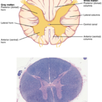
Cross-section of Spinal Cord
The cross-section of a thoracic spinal cord segment shows the posterior, anterior, and lateral horns of gray matter, as well as the posterior, anterior, and lateral columns of white matter
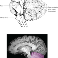
The Cerebellum
The cerebellum is situated on the posterior surface of the brain stem. Descending input from the cerebellum enters through the large white matter structure of the pons. Ascending input from the periphery and spinal cord enters through the fibers of…
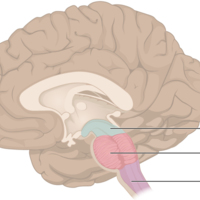
The Brain Stem
The brain stem comprises three regions: the midbrain, the pons, and the medulla.
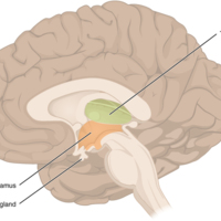
The Diencephalon
The diencephalon is composed primarily of the thalamus and hypothalamus, which together define the walls of the third ventricle. The thalami are two elongated, ovoid structures on either side of the midline that make contact in the middle. The…
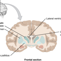
Frontal Section of Cerebral Cortex and Basal Nuclei
The major components of the basal nuclei, shown in a frontal section of the brain, are the caudate (just lateral to the lateral ventricle), the putamen (inferior to the caudate and separated by the large white-matter structure called the internal…
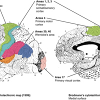
Brodmann's Areas of the Cerebral Cortex
Brodmann mapping of functionally distinct regions of the cortex was based on its cytoarchitecture at a microscopic level.
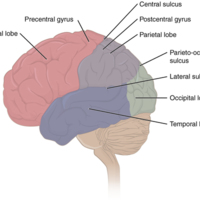
Lobes of the Cerebral Cortex
The cerebral cortex is divided into four lobes. Extensive folding increases the surface area available for cerebral functions.
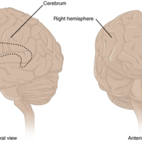
The Cerebrum
The cerebrum is a large component of the CNS in humans, and the most obvious aspect of it is the folded surface called the cerebral cortex.
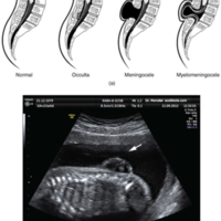
Spinal Bifida
(a) Spina bifida is a birth defect of the spinal cord caused when the neural tube does not completely close, but the rest of development continues. The result is the emergence of meninges and neural tissue through the vertebral column. (b) Fetal…
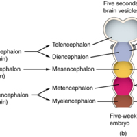
Primary and Secondary Vesicle Stages of Development
The embryonic brain develops complexity through enlargements of the neural tube called vesicles; (a) The primary vesicle stage has three regions, and (b) the secondary vesicle stage has five regions.
Receptor Types
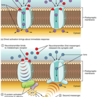
(a) An ionotropic receptor is a channel that opens when the neurotransmitter binds to it. (b) A metabotropic receptor is a complex that causes metabolic changes in the cell when the neurotransmitter binds to it (1). After binding, the G protein…
Leakage Channels
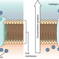
In certain situations, ions need to move across the membrane randomly. The particular electrical properties of certain cells are modified by the presence of this type of channel.
Voltage-Gated Channels
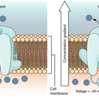
Voltage-gated channels open when the transmembrane voltage changes around them. Amino acids in the structure of the protein are sensitive to charge and cause the pore to open to the selected ion.
Testing the Water
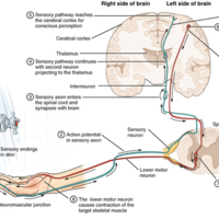
(1) The sensory neuron has endings in the skin that sense a stimulus such as water temperature. The strength of the signal that starts here is dependent on the strength of the stimulus. (2) The graded potential from the sensory endings, if strong…
The Process of Myelination
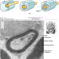
Myelinating glia wrap several layers of cell membrane around the cell membrane of an axon segment. A single Schwann cell insulates a segment of a peripheral nerve, whereas in the CNS, an oligodendrocyte may provide insulation for a few separate axon…
Glial Cells of the CNS
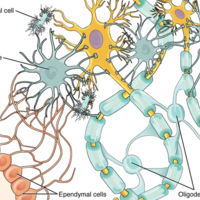
The CNS has astrocytes, oligodendrocytes, microglia, and ependymal cells that support the neurons of the CNS in several ways.
Parts of a Neuron
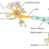
The major parts of the neuron are labeled on a multipolar neuron from the CNS.
Optic Nerve Versus Optic Tract
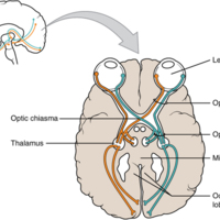
This drawing of the connections of the eye to the brain shows the optic nerve extending from the eye to the chiasm, where the structure continues as the optic tract. The same axons extend from the eye to the brain through these two bundles of fibers,…
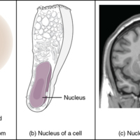
What Is a Nucleus
(a) The nucleus of an atom contains its protons and neutrons. (b) The nucleus of a cell is the organelle that contains DNA. (c) A nucleus in the CNS is a localized center of function with the cell bodies of several neurons, shown here circled in red
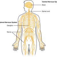
Central and Peripheral Nervous System
The structures of the PNS are referred to as ganglia and nerves, which can be seen as distinct structures. The equivalent structures in the CNS are not obvious from this overall perspective and are best examined in prepared tissue under the…
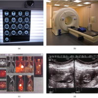
Medical Imaging Techniques
(a) The results of a CT scan of the head are shown as successive transverse sections. (b) An MRI machine generates a magnetic field around a patient. (c) PET scans use radiopharmaceuticals to create images of active blood flow and physiologic…
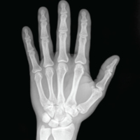
X-Ray of a Hand
High energy electromagnetic radiation allows the internal structures of the body, such as bones, to be seen in X-rays like these
Tags: Medicine & Health, X-Ray
Nursing Care at the End of Life: What Every Clinician Should Know
Nursing Care at the End of Life: What Every Clinician Should Know should be an essential component of basic educational preparation for the professional registered nurse student. Recent studies show that only one in four nurses feel confident in…
Malarial Subjects
Malaria was considered one of the most widespread disease-causing entities in the nineteenth century. It was associated with a variety of frailties far beyond fevers, ranging from idiocy to impotence. And yet, it was not a self-contained category.…
Tags: Malarial, Medicine & Health
Public Health Ethics: Cases Spanning the Globe
This Open Access book highlights the ethical issues and dilemmas that arise in the practice of public health. It is also a tool to support instruction, debate, and dialogue regarding public health ethics. Although the practice of public health has…
Tags: Medicine & Health, Public Health
Field Trials of Health Interventions: A Toolbox
Abstract
Before new interventions are released into disease control programmes, it is essential that they are carefully evaluated in `field trials'. These may be complex and expensive undertakings, requiring the follow-up of hundreds, or…
Before new interventions are released into disease control programmes, it is essential that they are carefully evaluated in `field trials'. These may be complex and expensive undertakings, requiring the follow-up of hundreds, or…
Diseases of Children in the Subtropics and Tropics
Paediatrics is often thought of as following two main routes. One is that of ultra-technology and ever-narrower specialization. The other is recognition of child health in a community context, related to family circumstances (especially the health…


